The term Abdo Pelvis is widely used in modern medical practice, especially in diagnostic radiology, to describe imaging of the abdomen and pelvis. Whether it is through a CT scan, MRI, ultrasound, or X-ray, an Abdo Pelvis scan plays a vital role in identifying, monitoring, and treating a wide range of health conditions. Doctors often recommend an Abdo Pelvis scan when patients present with abdominal pain, pelvic pain, trauma, or suspected internal disease. It helps specialists visualize critical abdominal cavity organs such as the liver, kidneys, spleen, pancreas, stomach, intestines, and also key pelvic cavity anatomy including the bladder, uterus, ovaries, prostate, and rectum.
An Abdo Pelvis scan is not just about looking inside the body—it is about providing patients with clarity, reassurance, and answers during uncertain times. By combining advanced cross-sectional imaging techniques with expert radiology interpretation, physicians can diagnose conditions early, guide treatment, and improve outcomes. The importance of Abdo Pelvis imaging cannot be overstated, as it is central to modern healthcare and trusted across the world.
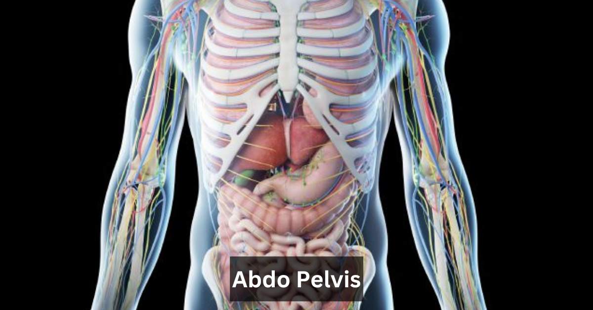
Understanding the Abdo Pelvis region begins with appreciating the complexity of the organs within it. The abdomen contains essential structures such as the liver, which regulates metabolism, the kidneys, which filter blood and maintain fluid balance, the spleen, which supports immunity, and the pancreas, responsible for insulin production and digestion. The stomach and intestines—including the small intestine, large intestine, and colon—are vital for nutrient absorption and waste elimination. Each organ plays a critical role in daily life, and imaging these areas provides insight into both acute and chronic conditions.
The pelvis houses equally important organs. In men, the prostate supports reproductive health, while in women, the uterus and ovaries are central to fertility and hormone regulation. The bladder stores urine, and the rectum forms the final part of the digestive system. In addition, major blood vessels such as the aorta and iliac arteries run through the Abdo Pelvis, supplying life-sustaining circulation to the body. Because of this complexity, accurate and detailed abdominal and pelvic imaging is essential for proper medical care.
Types of Abdo Pelvis Imaging
CT Abdo Pelvis
One of the most common techniques is the CT Abdo Pelvis scan. Using computed tomography, this method creates detailed cross-sectional imaging of the abdomen and pelvis. A CT Abdo Pelvis with contrast involves the use of a special contrast dye—often iodine-based—administered orally or intravenously, which highlights blood vessels, organs, and abnormal tissue. A CT Abdo Pelvis without contrast, on the other hand, is preferred in cases such as kidney stone detection or when contrast is contraindicated.
Doctors rely on CT Abdo Pelvis scans to detect appendicitis, bowel obstruction, kidney stones, abdominal trauma, and even hidden tumors. In emergencies, an emergency CT Abdo Pelvis can be lifesaving, helping doctors make rapid decisions about surgery or other interventions. A normal Abdo Pelvis CT will show clear organ structures without swelling, blockages, or masses, while abnormal Abdo Pelvis findings might reveal infection, inflammation, or cancer.
MRI Abdo Pelvis
An Abdo Pelvis MRI is another advanced imaging option, using strong magnets and radio waves rather than radiation. MRI excels at visualizing soft tissues, making it especially useful in detecting gynecological conditions such as ovarian cysts, uterine fibroids, and pelvic inflammatory disease, as well as tumors in the prostate and rectum. MRI is often chosen when gastrointestinal imaging requires precise detail or when patients need an alternative to radiation.
Doctors prefer MRI over CT when evaluating soft tissue changes, monitoring cancer progression, or avoiding radiation exposure, especially in younger patients. While the procedure may take longer, it provides an unparalleled level of clarity in certain conditions.
Ultrasound Abdo Pelvis
An Abdo Pelvis ultrasound is often the first imaging choice for patients with lower abdominal or pelvic pain. Using high-frequency sound waves, ultrasound is safe, non-invasive, and radiation-free. It is commonly performed for pregnancy scans, detection of gallstones, and evaluation of gynecological or urinary issues such as ovarian cysts, bladder tumors, or kidney problems. A ultrasound of abdomen and pelvis can quickly rule out many issues and is widely accessible, making it a cornerstone of diagnostic radiology.
X-ray Abdo Pelvis
While less detailed than CT or MRI, an X-ray Abdo Pelvis is still useful in specific situations, particularly trauma cases. It helps doctors assess fractures, dislocations, or certain obstructions. Though it is not typically the first choice for soft tissue evaluation, an X-ray provides quick, affordable, and widely available insights in urgent scenarios.
Why Doctors Order an Abdo Pelvis Scan
Doctors recommend an Abdo Pelvis scan for multiple reasons, each tied to improving patient outcomes. Patients with persistent abdominal or pelvic pain may need imaging to rule out serious issues like appendicitis, bowel obstruction, or urinary problems. In trauma situations, an Abdo Pelvis CT is often ordered immediately to identify internal bleeding or organ rupture.
Another reason is cancer detection and staging. Abdo Pelvis imaging is critical in spotting colorectal cancer, ovarian cancer, prostate cancer, and liver cancer. By providing detailed images, doctors can determine tumor size, spread, and treatment response. Additionally, Abdo Pelvis scans are used to monitor chronic conditions such as kidney disease, hernias, or inflammatory bowel disease, as well as for pre- and post-surgical imaging to guide medical teams during recovery.
Preparation for an Abdo Pelvis Scan
CT Scan Preparation
Before a CT Abdo Pelvis scan, patients may be instructed to fast for several hours. If contrast dye is used, hydration is encouraged to protect the kidneys. Patients should inform doctors of allergies or pre-existing kidney issues, as these may affect contrast use.
MRI Preparation
Preparation for an MRI Abdo Pelvis involves ensuring there are no metallic implants such as pacemakers, as these can interfere with the magnetic field. Patients may also be screened for claustrophobia, as MRI scans take longer and require stillness inside the scanner.
Ultrasound Preparation
For an Abdo Pelvis ultrasound, patients are often advised to drink water and maintain a full bladder. This enhances visibility of pelvic organs and provides clearer imaging. Unlike CT and MRI, ultrasound requires no fasting or injections, making it one of the simplest procedures for patients.
What to Expect During the Procedure
A CT Abdo Pelvis scan is quick and painless. Patients lie on a table that slides into the scanner while the machine takes images. If contrast is used, a warm sensation may occur when the dye is injected.
In an MRI Abdo Pelvis, the patient lies still in a cylindrical scanner while radio waves capture detailed images. The process takes longer than CT, but is also painless.
For an ultrasound Abdo Pelvis, a technician applies gel to the skin and moves a probe across the abdomen and pelvis. The procedure is comfortable, non-invasive, and produces immediate results.
Risks and Safety Considerations
CT scans involve radiation exposure, but the benefits often outweigh the risks, especially in emergencies. Allergic reactions to contrast dye are rare but possible.
MRI carries no radiation risk but is unsuitable for patients with certain implants. Ultrasound is considered the safest option, particularly for pregnant women and children.
Interpreting Abdo Pelvis Results
A normal Abdo Pelvis report will show clear, well-defined organs with no signs of disease. An abnormal Abdo Pelvis finding may include inflammation, blockages, tumors, or fluid collections.
Radiologists interpret the scans using advanced tools such as PACS and DICOM imaging files, ensuring accurate results are shared with doctors. These interpretations are critical in guiding treatment decisions.
Conditions Diagnosed with Abdo Pelvis Imaging
Digestive System Conditions
Abdo Pelvis scans can detect appendicitis, bowel obstruction, and tumors of the stomach or intestines. Early detection through imaging reduces complications and speeds up treatment.
Urinary and Kidney Conditions
An Abdo Pelvis CT or ultrasound is ideal for spotting kidney stones, urinary tract obstructions, and bladder tumors. These scans help guide both surgical and non-surgical management.
Gynecological & Reproductive Conditions
For women, an Abdo Pelvis MRI or ultrasound can detect ovarian cysts, uterine fibroids, and pelvic inflammatory disease. These conditions are often managed more effectively with early diagnosis.
Male Pelvic Conditions
Men benefit from Abdo Pelvis imaging for conditions like prostate enlargement, prostate cancer, and testicular abnormalities. MRI in particular offers excellent detail of soft tissue in these cases.
Trauma and Emergency Cases
In trauma, an Abdo Pelvis scan can reveal internal bleeding, organ rupture, and fractures, making it a crucial tool in saving lives after accidents or injuries.
Cost and Availability of Abdo Pelvis Scans
The cost of an Abdo Pelvis scan varies depending on whether it is CT, MRI, or ultrasound. CT is generally more affordable than MRI, while ultrasound is usually the least expensive. Availability is widespread in hospitals and diagnostic centers, and many insurance plans cover part or all of the cost.
Alternatives to Abdo Pelvis Imaging
While Abdo Pelvis imaging is highly effective, other diagnostic tools may also be used. Blood tests, urine tests, and endoscopy can provide supporting information. In some cases, a PET scan may be used alongside CT to assess cancer spread.
Conclusion
The Abdo Pelvis scan is one of the most valuable tools in modern medicine, offering safe, detailed, and reliable insight into the most vital organs of the human body. Whether performed as a CT Abdo Pelvis with contrast, an Abdo Pelvis MRI, or an ultrasound Abdo Pelvis, this diagnostic test provides clarity and peace of mind. With its ability to detect life-threatening conditions like appendicitis, kidney stones, or cancer, Abdo Pelvis imaging is a powerful ally in healthcare.
For patients, understanding what an Abdo Pelvis scan involves helps reduce anxiety and builds trust in the process. With expert radiologists, advanced imaging technologies, and supportive care teams, these scans empower both patients and doctors to take confident steps toward better health.
Frequently Asked Questions
Is an Abdo Pelvis scan painful?
No, all imaging methods are painless and safe.
How long does it take?
CT takes minutes, MRI takes 30–60 minutes, ultrasound takes 15–30 minutes.
Do I need a referral?
Yes, imaging usually requires a doctor’s referral.
Ultrasound is safe; CT and MRI are used only when necessary.
Ultrasound is safe; CT and MRI are used only when necessary.
How soon will I get my results?
Reports are often available within 24–48 hours.
Stay in touch to get more updates & alerts on Coop Magazine! Thank you